 Examining the changes in the spine curve during growth is of great importance in early identification and prevention of the development of structural disorders of the spine. However, most of the studies published in this regard have focused on the use of X-rays as the gold standard for spine evaluation. Therefore, considering the ethical aspects of the widespread use of harmful X-rays, few studies can be found that have investigated the normal values of the kyphosis angle, especially in children without a background of disease and spine disorders, by conducting an evaluation in a large population.
Normal values of the kyphosis angle:
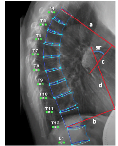
Using x-rays, the angle difference between the first and twelfth thoracic vertebrae is usually measured as the kyphosis angle. This value is usually between 20 and 40 degrees in the young population, between 48 and 50 degrees in elderly women, and around 44 degrees in elderly men. Values above 50 degrees are classified as hyperkyphosis.
Normal values of the kyphosis angle in growing age:
Considering the mentioned limitations in the widespread use of X-rays, the article by Giglio et al. in the Journal of Children’s Orthopaedics is one of the few references of studies conducted on a large number of healthy people in the growing age. In this article, which was published in 2007, 718 study cases in the age of 5 to 20 years have been evaluated using a precise laboratory instrument.
At first, the laboratory measurement method was validated by comparing the results of 20 healthy individuals with the results obtained from radiography, and after ensuring the accuracy of the results, 718 healthy cases, including 350 boys and 368 girls, were evaluated in terms of kyphosis angles. The results show a linear increase in the measured values from 25 degrees at the age of 7 years to 38 degrees at the age of 19 years. Finally, the researchers of this study suggested the relationship Kyphotic Angle=25+0.58*age as a suitable linear curve to identify the normal value of the kyphosis angle in growing children, regardless of their gender.
Using spinal mouse to measure kyphosis:
The use of alternative systems to reduce the risks caused by X-rays has led to the development of new technologies in the evaluation of the spine. Spinal mouse systems can be mentioned among these technologies. The Spinal Mouse device is a system consisting of precise motion sensors that allows the therapist to accurately evaluate its three-dimensional curves by moving on the spine. Spinal Mouse, which does not have any side effects caused by harmful rays, has been evaluated in several studies in the evaluation of kyphosis, lordosis and scoliosis angles, and due to the high validity and reliability reported in the results, it is now widely used in centers. Research and treatment are being used all over the world.